CT Angiography and Your Kidneys: Understanding the Risks
CT angiography uses iodinated contrast that can harm kidneys in at-risk patients. Learn who is most at risk, how it's prevented, and how to stay safe.
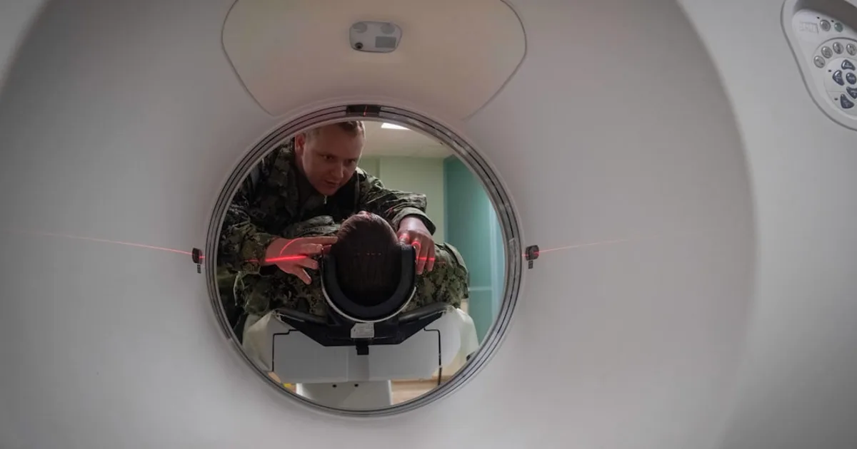
What is CT angiography? CT angiography (CTA) is an imaging technique that uses X-rays and an intravenous injection of iodinated contrast medium to visualise blood vessels in detail — arteries, veins, and the heart. It is widely used to assess conditions such as aortic aneurysm, peripheral artery disease, and carotid stenosis. Contrast-induced nephropathy (CIN), also called contrast-associated acute kidney injury (CA-AKI), refers to a deterioration in kidney function occurring within 48–72 hours of iodinated contrast administration, in the absence of another cause [4].
CT angiography has transformed vascular medicine. In a few minutes, it can map the entire arterial tree of the body, detect a dangerous aneurysm, or identify the location of a blockage threatening a limb. But this powerful tool has a well-known limitation: iodinated contrast medium, the substance that makes vessels visible on the scan, can damage the kidneys.
For the vast majority of patients, this risk is small and manageable. But for those with pre-existing kidney disease, diabetes, or dehydration, it requires genuine attention — and a clear conversation with the medical team before the procedure.
How common is contrast-induced kidney injury?
Pooled data across studies suggest that contrast-induced kidney injury occurs in approximately 6% of patients receiving contrast-enhanced CT scans [3]. This figure, however, masks enormous variation depending on the patient’s baseline kidney function.
In a large meta-analysis of 40 studies, Kooiman et al. found that the risk of a significant rise in creatinine after contrast CT was approximately 6%, while the risk of dialysis was extremely low at 0.06%, and persistent kidney function decline occurred in about 1% of affected patients [3].
A separate cohort study found that, in outpatients attending for routine contrast-enhanced CT, the incidence of CIN reached 11% — and it was linked to increased risk of severe renal failure and death [5].
To put this in context: a patient with normal kidneys undergoing a single elective CT angiogram faces a very low risk. A patient with advanced chronic kidney disease (eGFR below 30), diabetes, and dehydration faces a genuinely elevated risk that warrants specific precautions.
It is worth noting that some recent analyses have questioned whether the risk is being overestimated. A 2018 review found no significant increase in acute kidney injury or mortality when comparing contrast-enhanced CT to non-contrast CT [8]. This nuance matters: CIN may sometimes reflect the natural progression of underlying kidney disease rather than a direct causal effect of contrast.
Who is most at risk?
Several factors are consistently associated with higher risk of contrast-induced kidney injury [3][4][6]:
Kidney function (the main determinant)
| eGFR (mL/min/1.73m²) | Approximate CIN risk |
|---|---|
| Above 60 | Very low |
| 45–59 | Low (~0%) |
| 30–44 | Moderate (~2.9%) |
| Below 30 | High (~12.1%) |
Diabetes mellitus Diabetes approximately triples the risk of contrast-induced kidney injury, with an odds ratio of 3.10 in meta-analyses [3]. This is particularly relevant in patients with both diabetes and reduced eGFR.
Dehydration Any state of reduced blood volume — whether from vomiting, diarrhoea, diuretics, or simply insufficient fluid intake — concentrates contrast in the kidney tubules and amplifies its toxic effects.
Concurrent nephrotoxic drugs Non-steroidal anti-inflammatory drugs (NSAIDs), aminoglycoside antibiotics, and certain other medications impair the kidney’s ability to manage contrast toxicity. Some of these are temporarily paused before high-risk procedures.
Volume of contrast administered Larger volumes of contrast mean greater exposure. This is why CT angiography — which typically requires more contrast than standard CT for the same patient — carries slightly higher renal risk [1][4].
Age and cardiovascular comorbidities Advanced age, heart failure, and reduced cardiac output reduce renal perfusion and are compounding risk factors [4].
What happens when kidneys are injured by contrast?
In most cases, contrast-induced kidney injury is a transient, self-resolving rise in serum creatinine, detected on blood tests 48–72 hours after the scan and resolving within a week without treatment.
In a small proportion of cases, however, the consequences can be more serious:
- Persistent kidney function decline: approximately 1.1% of affected patients sustain lasting damage [3]
- Need for dialysis: rare but documented, occurring in approximately 0.06% of all patients receiving contrast [3]
- Increased mortality: studies have shown an association between CIN and increased in-hospital and long-term mortality, though the extent to which CIN itself — versus the underlying illness — drives this association remains debated [4][5]
- Poor long-term kidney survival: patients with eGFR below 30 who develop CIN face particularly poor long-term renal outcomes [6]
A study in cerebrovascular patients undergoing CT angiography and CT perfusion imaging found that in only 0.37% of 1,075 patients did contrast possibly contribute to renal failure, with 0.19% requiring temporary haemodialysis — demonstrating that even in this intensive neurological context, the absolute risk was modest [1].
How does contrast damage the kidneys?
The mechanisms are multiple and partially overlapping [7]:
Renal medullary hypoxia Iodinated contrast causes vasoconstriction of the small vessels supplying the kidney’s medulla (inner zone). This reduces oxygen delivery to tubular cells, which are already working in a low-oxygen environment and are particularly vulnerable.
Direct cellular toxicity Contrast molecules have a direct cytotoxic effect on renal tubular cells, disrupting cellular membranes and mitochondrial function.
Oxidative stress Contrast administration triggers the production of reactive oxygen species (free radicals), which cause oxidative damage to tubular cells.
Apoptosis and inflammation Tubular cell death via programmed apoptosis, and subsequent inflammatory cascades, amplify the initial injury and delay recovery.
Prevention: what actually works?
1. Intravenous hydration — the cornerstone
The single most effective preventive measure is IV isotonic saline (0.9% NaCl), administered before and after the procedure. Hydration increases renal blood flow, dilutes the contrast in tubular fluid, and reduces the duration of tubular exposure. This is the standard of care for all patients identified as at risk [4][6].
In the outpatient elective setting, this typically means IV saline at 1 mL/kg/hour for 6–12 hours before the scan and 4–6 hours afterwards. In emergency settings, the approach is adapted to clinical urgency.
2. Low-osmolar and iso-osmolar contrast agents
Modern contrast agents are far safer than their predecessors. Low-osmolar (e.g. iopromide, iohexol) and iso-osmolar (e.g. iodixanol) contrast media are significantly less nephrotoxic than the older high-osmolar agents, which are no longer in routine use [4].
3. Minimising contrast volume
The less contrast used, the lower the renal exposure. Radiologists adjust protocols to use the minimum volume compatible with diagnostic quality, particularly in high-risk patients [4].
4. Withholding nephrotoxic drugs temporarily
NSAIDs, aminoglycosides, and diuretics may be paused for 24–48 hours before a high-risk procedure. Metformin is typically paused after — not before — contrast administration in patients with impaired kidney function, due to the theoretical risk of lactic acidosis if kidney function deteriorates [4].
5. N-acetylcysteine (NAC) — no longer recommended
For many years, oral NAC was widely prescribed before contrast procedures. However, the landmark PRESERVE trial (n=4,993, Weisbord et al., NEJM 2018, PMID 29091581) definitively demonstrated that NAC is no better than placebo and sodium bicarbonate no better than isotonic saline. Current guidelines do not recommend NAC for routine CIN prevention.
6. Statins — emerging evidence
High-dose statins (rosuvastatin, atorvastatin) have shown some promise in reducing CIN incidence, particularly in patients with diabetes undergoing cardiac catheterisation. The mechanism may involve anti-inflammatory and antioxidant effects [7]. Evidence in the CT context specifically is less robust, and statins should not be started solely for this purpose.
Weighing benefits against risks
The key clinical decision is not “is there a risk?” but rather “does the benefit of the scan outweigh the risk for this patient?”
In emergencies — suspected aortic dissection, acute limb ischaemia, stroke — the diagnostic information from CT angiography can be lifesaving. The risk of withholding the scan far outweighs the renal risk of the contrast. No hesitation is warranted.
In elective situations — pre-operative planning, surveillance of known disease — there is time to assess renal function, optimise hydration, and consider alternatives:
- Ultrasound (Doppler): no contrast, no radiation — first-line for many vascular assessments
- MRI angiography (MRA): uses gadolinium contrast or can sometimes be performed without contrast. In very advanced renal failure (eGFR below 15–20), gadolinium carries its own specific risk (nephrogenic systemic fibrosis)
- CO₂ angiography: a contrast-free catheter-based technique used in some centres for patients with severe CKD
- Risk stratification tools: the Mehran risk score [4] integrates eGFR, diabetes, haemodynamic status, and planned contrast volume to stratify individual risk
For patients with known peripheral artery disease or abdominal aortic aneurysm requiring CT angiography for surveillance or treatment planning, kidney function should be checked before each study.
CT angiography: a specific challenge
Standard contrast CT (e.g. chest CT for pulmonary embolism) typically uses 50–100 mL of contrast. CT angiography for vascular assessment of the aorta or lower-limb arteries may require 100–150 mL — sometimes more in complex multi-territory studies.
This higher contrast volume, combined with the fact that vascular patients frequently have coexisting risk factors (age, diabetes, atherosclerosis affecting renal arteries), means that renal risk assessment is particularly important in the CTA context [1][4].
Modern CT technology — faster scanners, better detector sensitivity — is progressively reducing the contrast volumes required for diagnostic quality, which is good news for patients with fragile kidneys.
Practical checklist before your CT angiogram
If you have been referred for a CT angiogram and have kidney disease, here is what to discuss with your doctor:
- Have your kidney function tested — blood creatinine and eGFR before the procedure
- Stay well hydrated in the days before — avoid being fasted for longer than necessary
- List all your medications — some may need temporary adjustment (NSAIDs, diuretics, metformin)
- Tell your radiologist and GP if you have diabetes, CKD, or a family history of kidney disease
- Ask about IV hydration — if you are at risk, it should be offered as standard
- Follow up with a blood test 48–72 hours after the scan if your doctor advises it
Conclusion
CT angiography is an essential tool in vascular medicine — it saves lives. The risk to kidneys is real but well understood, and for most patients it is either negligible or effectively managed with straightforward precautions. The key is identifying at-risk patients before the scan, applying protective measures systematically, and weighing the diagnostic benefit honestly against the renal risk.
If you have kidney disease or diabetes and have been referred for a CT angiogram, do not refuse the scan — but do have the conversation with your doctor. The information obtained from the scan, and the way it guides your treatment, is almost always worth the managed risk.
This article is for informational purposes only and does not constitute medical advice. Always consult your GP or a vascular specialist or nephrologist for decisions regarding your health.
References
- Josephson SA, Dillon WP, Smith WS. Incidence of contrast nephropathy from cerebral CT angiography and CT perfusion imaging. Neurology. 2005;64(10):1805-1806. PMID 15911820. DOI: 10.1212/01.wnl.0000161845.69114.62
- Garfinkle MA, Stewart S, Basi R. Incidence of CT contrast agent-induced nephropathy: toward a more accurate estimation. AJR Am J Roentgenol. 2015;204(6):1146-1151. PMID 26001222. DOI: 10.2214/ajr.14.13761
- Kooiman J, Pasha SM, Zondag W, et al. Meta-analysis: serum creatinine changes following contrast enhanced CT imaging. Eur J Radiol. 2012;81(10):2554-2561. PMID 22177326. DOI: 10.1016/j.ejrad.2011.11.020
- Mehran R, Dangas GD, Weisbord SD. Contrast-associated acute kidney injury. N Engl J Med. 2019;380(22):2146-2155. PMID 31141635. DOI: 10.1056/nejmra1805256
- Mitchell AM, Jones AE, Tumlin JA, Kline JA. Incidence of contrast-induced nephropathy after contrast-enhanced computed tomography in the outpatient setting. Clin J Am Soc Nephrol. 2010;5(1):4-9. PMID 19965528. Free full text: PMC2801649. DOI: 10.2215/cjn.05200709
- Kim SM, Cha RH, Lee JP, et al. Incidence and outcomes of contrast-induced nephropathy after computed tomography in patients with CKD. Am J Kidney Dis. 2010;55(6):1018-1025. PMID 20097462. DOI: 10.1053/j.ajkd.2009.10.057
- Zhang F, Lu Z, Wang F. Advances in the pathogenesis and prevention of contrast-induced nephropathy. Life Sci. 2020;259:118379. PMID 32890604. DOI: 10.1016/j.lfs.2020.118379
- Patel T, Patel V. Review: in adults, contrast-enhanced CT is not linked to acute kidney injury or mortality vs noncontrast CT. Ann Intern Med. 2018;168(2):JC10. PMID 29335722. DOI: 10.7326/acpjc-2018-168-2-010
- Weisbord SD, Gallagher M, Jneid H, et al. Outcomes after angiography with sodium bicarbonate and acetylcysteine. N Engl J Med. 2018;378(7):603-614. PMID 29091581. DOI: 10.1056/NEJMoa1710933 — PRESERVE trial: NAC no better than placebo for CIN prevention.
Frequently asked questions
Can I have a CT angiogram if I have chronic kidney disease?
What symptoms should I watch for after a contrast CT scan?
Does drinking lots of water before the scan protect my kidneys?
Is CT angiography riskier for the kidneys than a standard contrast CT?
Petit Veinard Editorial Board
This article was written and reviewed by vascular medicine specialists. Sources: peer-reviewed journals (PubMed), ESVS guidelines, AHA/ACC recommendations, Cochrane Reviews.