Chronic Venous Insufficiency: Symptoms, Stages, and Treatment
CVI affects millions worldwide. Learn its CEAP stages, warning signs, diagnosis, and treatments — with evidence from European and American guidelines.
Citable definition: Chronic venous insufficiency (CVI) is a progressive vascular condition in which the veins of the lower limbs fail to return blood efficiently to the heart due to valvular incompetence (leaky one-way valves), venous obstruction, or both — leading to sustained elevated venous pressure, tissue changes, and, in advanced cases, non-healing ulcers. It is classified internationally using the CEAP system (Clinical, Etiologic, Anatomic, Pathophysiologic), with clinical stages ranging from C0 to C6 (Kistner et al., Mayo Clinic Proceedings, 1996, PMID: 8637255; Lurie et al., J Vasc Surg Venous Lymphat Disord, 2020, PMID: 32113854).
What Is Chronic Venous Insufficiency?
Chronic venous insufficiency (CVI) is one of the most prevalent vascular conditions in the developed world, yet it is frequently underestimated — dismissed as a cosmetic inconvenience rather than recognized as the progressive, systemic condition it can become.
Under normal circumstances, a series of one-way valves inside the leg veins prevent blood from flowing backward as it travels upward against gravity toward the heart. In CVI, these valves become damaged or incompetent (unable to close properly), allowing blood to pool in the lower limbs — a phenomenon called venous reflux (backward blood flow). In some patients, the problem is not valve failure but venous obstruction (a partial or complete blockage of the vein), or a combination of both.
The result is chronically elevated pressure within the veins of the leg — a state called venous hypertension (persistently raised blood pressure inside the veins). Over time, this increased pressure damages the surrounding tissues, causing inflammation, skin changes, and, in the most severe cases, open wounds known as venous leg ulcers.
Importantly, CVI is not merely a local leg problem. A large population study published in the European Heart Journal found a significant association between CVI, cardiovascular disease, and increased overall mortality in the general population, suggesting that CVI should be viewed as a systemic risk marker rather than an isolated benign condition (Prochaska et al., Eur Heart J, 2021, PMID: 34387673).
For a broader overview of venous conditions, visit our veins section.
The CEAP Classification: Staging the Disease
The international language of CVI is the CEAP classification — an acronym standing for Clinical, Etiologic, Anatomic, and Pathophysiologic. First proposed in 1996 (Kistner et al., Mayo Clin Proc, 1996, PMID: 8637255), it was substantially revised in 2020 to improve diagnostic precision and inter-observer reproducibility (Lurie et al., J Vasc Surg Venous Lymphat Disord, 2020, PMID: 32113854).
The clinical dimension — the “C” in CEAP — is the most visible to patients:
| CEAP Stage | Description |
|---|---|
| C0 | No visible or palpable signs of venous disease |
| C1 | Spider veins (telangiectasias) or reticular veins |
| C2 | Varicose veins (≥3 mm diameter); C2s = symptomatic, C2r = recurrent |
| C3 | Edema (swelling) without skin changes |
| C4a | Pigmentation or eczema |
| C4b | Lipodermatosclerosis (hardening and discoloration of skin) or atrophie blanche (white scarring patches) |
| C4c | Corona phlebectatica (fan-shaped small veins at the foot) — added in 2020 |
| C5 | Healed venous ulcer |
| C6 | Active venous ulcer; C6r = recurrent active ulcer |
The 2020 revision introduced important refinements, including the new C4c subcategory and improved reporting standards for recurrent and symptomatic disease, making the system more clinically actionable (Lurie et al., PMID: 32113854).
Symptoms: What Does CVI Feel Like?
Symptoms vary considerably depending on the CEAP stage. They typically worsen throughout the day — particularly after prolonged standing or sitting — and improve with leg elevation.
Early-stage symptoms (C0–C2):
- A feeling of heaviness, fatigue, or aching in the legs
- Itching or burning around visible veins
- Visible spider veins or varicose veins
- Mild ankle swelling at the end of the day
Intermediate-stage symptoms (C3–C4):
- Persistent ankle and lower-leg edema (swelling)
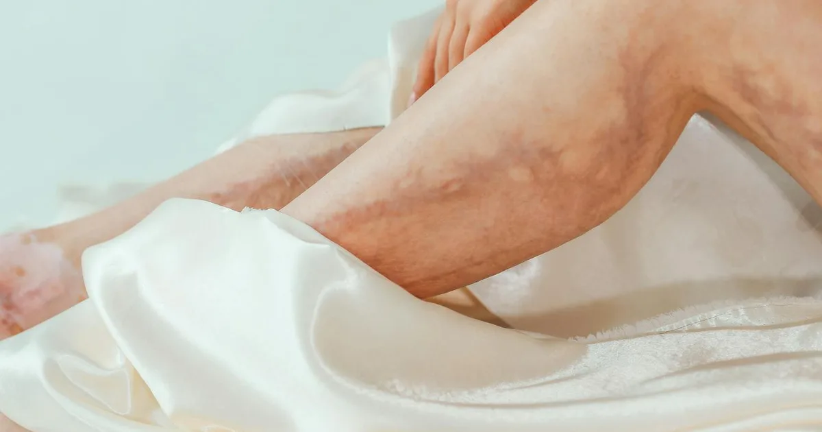
- Skin discoloration — brownish patches (hemosiderin deposits from leaked red blood cells)
- Dry, itchy, inflamed skin (venous eczema)
- Hardening of the skin and subcutaneous tissue (lipodermatosclerosis)
Advanced-stage symptoms (C5–C6):
- Non-healing or slowly healing wounds, typically around the inner ankle
- Wound pain, exudate (fluid discharge), and susceptibility to infection
- Significant impact on mobility and quality of life
Research published in 2026 confirms that patients who develop venous leg ulcers (C6) have significantly worse long-term survival compared to CVI patients without ulcers — making the ulcer stage a serious, independent prognostic marker (Chervonski et al., Phlebology, 2026, PMID: 41717669).
Diagnosis: What to Expect
Clinical Examination
A vascular specialist will begin with a thorough history and physical examination, assessing the distribution of varicose veins, skin changes, and the presence of edema. The CEAP classification guides this structured assessment (Rutherford Vascular Surgery, Chapter 19).
Duplex Ultrasound
Duplex ultrasound (a combined imaging and blood-flow measurement technique) is the cornerstone of CVI diagnosis. It identifies the location and extent of venous reflux and obstruction in the superficial, deep, and perforating (connecting) veins. It is non-invasive, widely available, and highly informative (Rutherford Vascular Surgery, Vascular Laboratory chapter).
Physiological Assessment
For more complex cases, the vascular laboratory offers additional tools:
- Ambulatory venous pressure (AVP) measurement: The gold-standard physiological test, directly measuring the pressure inside a foot vein during walking. It quantifies the severity of venous dysfunction but is invasive and rarely used in routine practice.
- Air plethysmography (APG): A non-invasive technique using a pneumatic cuff to measure venous filling, calf muscle pump function, and reflux volume. Studies cited in Rutherford Vascular Surgery show strong agreement between APG and duplex ultrasound (κ = 0.83), making it a reliable complement to imaging.
- Photoplethysmography (PPG): A simpler optical technique that detects venous reflux with a sensitivity of 100%, but a specificity of only 60% — meaning it catches nearly all reflux cases but produces a significant number of false positives. The agreement with duplex is moderate (κ = 0.47), so it is better suited to screening than definitive diagnosis (Rutherford Vascular Surgery).
Treatment: From Conservative to Surgical
Treatment is tailored to CEAP stage, symptom burden, and the patient’s overall health. The goal is to reduce venous hypertension, relieve symptoms, prevent progression, and heal or prevent ulcers.
Conservative (Non-Interventional) Management
| Approach | Details |
|---|---|
| Compression therapy | Medical-grade compression stockings (class I–III) are the foundation of CVI management at all stages. They reduce venous hypertension and edema. |
| Leg elevation | Elevating legs above heart level for 30 minutes, 3–4 times per day, reduces venous pooling. |
| Physical activity | Walking activates the calf muscle pump — the body’s natural venous return mechanism. |
| Weight management | Excess body weight increases intra-abdominal pressure and impairs venous return. |
| Venoactive drugs | Micronized purified flavonoid fraction (MPFF) and other phlebotonic agents may reduce symptoms in early-to-intermediate stages; discuss options with your physician. |
Interventional and Surgical Options
For patients with significant reflux, persistent symptoms, or complications despite conservative measures, interventional treatment is indicated:
- Endovenous thermal ablation (laser or radiofrequency): Minimally invasive techniques that close incompetent superficial veins using heat delivered through a thin catheter. These are now the preferred first-line procedures in most European centers.
- Foam sclerotherapy (injection of a foam agent to close the vein): Suitable for smaller varicose veins and recurrent disease.
- Phlebectomy (surgical removal of varicose veins through tiny incisions): Often performed alongside ablation.
- Deep venous reconstruction: Reserved for complex cases with deep venous obstruction or severe valve incompetence, performed by specialist vascular surgeons.
- Venous leg ulcer care: Requires a multidisciplinary approach — compression bandaging, wound care, and often interventional treatment of the underlying reflux.
Explore our treatments section for in-depth articles on each of these procedures.
Prevention: Daily Habits That Make a Difference
While genetic predisposition plays a role in CVI, several modifiable risk factors are well established. Our editorial board recommends the following evidence-informed habits:
- Move regularly — avoid sitting or standing for more than 30–45 minutes without a short walk or calf-raise exercise.
- Wear compression hosiery if you have a family history of varicose veins, work in a sedentary or standing profession, or are pregnant.
- Maintain a healthy weight — even modest weight loss reduces venous pressure in the lower limbs.
- Elevate your legs in the evening, particularly after a long day on your feet.
- Stay hydrated — adequate hydration supports healthy blood viscosity.
- Avoid prolonged heat exposure (hot baths, saunas) during active disease flares, as heat causes venous dilation and worsens pooling.
- Do not smoke — smoking impairs microcirculation and wound healing, which is particularly critical at advanced CEAP stages.
For more prevention strategies, visit our prevention section.
When to See a Doctor
Do not wait for an open wound before seeking medical attention. Consult your physician or a vascular specialist if you notice:
- Persistent leg swelling that does not resolve overnight with elevation
- Skin discoloration or hardening around the lower leg or ankle
- A wound near the ankle that has not healed within two weeks
- Sudden, severe leg pain or swelling — this may indicate a deep vein thrombosis (DVT, a blood clot in a deep vein) and requires urgent evaluation
- Varicose veins that are painful, warm, or red — signs of superficial thrombophlebitis (inflammation and clotting in a surface vein)
- Any stage of CVI that is affecting your quality of life, sleep, or mobility
The evidence is clear: CVI is a progressive condition, and earlier intervention consistently leads to better outcomes. The development of a venous leg ulcer is associated with significantly reduced long-term survival (Chervonski et al., PMID: 41717669) — a compelling reason not to delay evaluation.
Sources
-
Chervonski E, Bisen SS, Jacobowitz GR, Rockman CB, Maldonado TS, et al. Venous leg ulcers are a marker of worse long-term survival in patients treated for chronic venous insufficiency. Phlebology. 2026 Feb 20. DOI: 10.1177/02683555261429077 — PMID: 41717669
-
Prochaska JH, Arnold N, Falcke A, Kopp S, Schulz A, Buch G, et al. Chronic venous insufficiency, cardiovascular disease, and mortality: a population study. Eur Heart J. 2021 Oct 21;42(40):4157–4165. DOI: 10.1093/eurheartj/ehab495 — PMID: 34387673
-
Kistner RL, Eklof B, Masuda EM. Diagnosis of chronic venous disease of the lower extremities: the “CEAP” classification. Mayo Clin Proc. 1996 Apr;71(4):338–45. DOI: 10.4065/71.4.338 — PMID: 8637255
-
Lurie F, Passman M, Meisner M, Dalsing M, Masuda E, Welch H, et al. The 2020 update of the CEAP classification system and reporting standards. J Vasc Surg Venous Lymphat Disord. 2020 May;8(3):342–352. DOI: 10.1016/j.jvsv.2019.12.075 — PMID: 32113854
-
Rutherford RB (ed.). Rutherford’s Vascular Surgery, 8th edition. Chapter 19: Clinical Evaluation of the Venous and Lymphatic Systems; Vascular Laboratory: Venous Physiologic Assessment. Elsevier Saunders. [No PMID — textbook reference]
Medical Disclaimer: This article is produced by the Petit Veinard Editorial Board for informational and educational purposes only. It does not constitute medical advice, diagnosis, or a treatment recommendation. The information presented reflects published scientific literature and international clinical guidelines available at the time of writing. Individual clinical situations vary; always consult a qualified physician or vascular specialist before making any decisions about your health. In case of acute symptoms — such as sudden leg swelling, chest pain, or shortness of breath — seek emergency medical care immediately.
Frequently asked questions
What are the first signs of chronic venous insufficiency in the legs?
Is chronic venous insufficiency dangerous or just cosmetic?
What is the CEAP classification and why does my doctor use it?
Petit Veinard Editorial Board
This article was written and reviewed by vascular medicine specialists. Sources: peer-reviewed journals (PubMed), ESVS guidelines, AHA/ACC recommendations, Cochrane Reviews.