Platelet-Rich Fibrin (PRF) for Chronic Venous Ulcer Healing
How PRF therapy accelerates chronic venous leg ulcer healing using your own blood platelets — latest 2026 clinical evidence explained.
Citable definition: Platelet-rich fibrin (PRF) is an autologous (derived from the patient’s own blood) biological scaffold rich in platelets, leukocytes (white blood cells), and growth factors, prepared by centrifuging whole blood without anticoagulants, and used as a wound-healing adjunct to accelerate tissue repair in chronic ulcers.
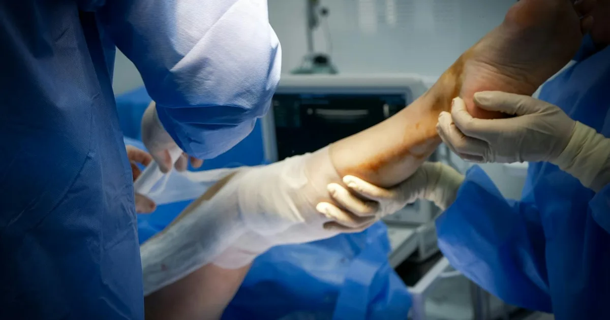
Chronic venous leg ulcers are among the most stubborn wounds in vascular medicine. They recur, they linger for months or years, and they impose a significant burden on patients’ quality of life. Standard compression therapy and wound dressings remain the cornerstone of treatment — but for ulcers that simply refuse to close, clinicians are increasingly turning to a technology derived from the patient’s own blood: platelet-rich fibrin (PRF).
This article explains what PRF is, how it works, what the latest clinical evidence says, and what patients with non-healing leg ulcers should know before asking their doctor about it.
What Is Platelet-Rich Fibrin?
PRF is a second-generation platelet concentrate. To prepare it, a small volume of the patient’s blood is drawn and placed in a centrifuge (a machine that spins samples at high speed to separate their components). Without the addition of any anticoagulants or chemical activators, the spinning separates the blood into layers. The middle layer — a golden, gel-like clot — is the PRF membrane. It contains a dense network of fibrin (the structural protein of blood clots), platelets, and leukocytes, all embedded with a cocktail of growth factors including PDGF (platelet-derived growth factor), TGF-β (transforming growth factor-beta), VEGF (vascular endothelial growth factor, which promotes new blood vessel formation), and IGF-1 (insulin-like growth factor 1).
Unlike its predecessor, platelet-rich plasma (PRP), PRF releases these growth factors gradually over several days rather than all at once. This slow, sustained release is thought to more closely mimic the body’s natural healing cascade.
Why does this matter for venous ulcers? Chronic venous leg ulcers develop when sustained high pressure in the leg veins (a condition called chronic venous insufficiency, or CVI) damages the skin and underlying tissue, creating wounds that lack the biological signals needed to heal normally. The local wound environment is often depleted of growth factors and dominated by inflammatory cells. PRF aims to correct this deficit by flooding the wound bed with the patient’s own regenerative proteins.
For a broader overview of venous conditions and how they affect the legs, see our vein health guide.
Symptoms of Chronic Venous Leg Ulcers
Before exploring PRF as a treatment, it is worth recognizing the typical presentation of the condition it targets:
- Open wound on the lower leg or ankle that has been present for more than four to six weeks (the standard clinical threshold for “chronic”)
- Irregular, shallow wound edges with a red or yellow wound bed
- Surrounding skin changes: brownish discoloration (called lipodermatosclerosis — hardening and darkening of the skin around the ankle due to chronic venous pressure), varicose eczema, or swelling
- Leg heaviness and aching, especially after prolonged standing
- Odor or discharge if the wound becomes colonized by bacteria
- Pain, which can range from mild to severe and is often worse at the end of the day
These ulcers account for approximately 70–80% of all chronic leg ulcers and affect an estimated 1–3% of the adult population in Western countries, with prevalence rising sharply in people over 65, according to available data.
How PRF Works: The Biology of Healing
Recent laboratory and animal research has begun to clarify the molecular pathways through which PRF promotes tissue repair. A 2025 study in rats with chronic refractory (treatment-resistant) wounds found that PRF accelerated healing by activating the Wnt/β-catenin signaling pathway — a molecular cascade central to cell proliferation, migration, and the formation of new connective tissue (Huang H, Huang M, Wang X, Yin G; Postępy Dermatologii i Alergologii, 2025; DOI: 10.5114/ada.2025.148762; PMID: 40521068). This provides a mechanistic rationale for the clinical benefits observed in human trials.
In parallel, materials scientists are exploring how PRF can be incorporated into novel delivery systems. A 2026 study published in the Journal of Materials Chemistry B described a PRF-loaded microneedle patch — a device with microscopic projections that deliver PRF directly into wound tissue for pressure injury repair — demonstrating that the growth factors retained their biological activity and promoted faster wound closure in preclinical models (Huang L et al.; J Mater Chem B, 2026; DOI: 10.1039/d5tb02111a; PMID: 41397385). While this technology is not yet in routine clinical use, it illustrates the direction of innovation in PRF delivery.
Clinical Evidence: What Do the Trials Say?
PRF vs. Normal Saline in Venous Leg Ulcers
The most directly relevant evidence for venous ulcer patients comes from a randomized controlled trial (RCT — the gold standard of clinical research) published in Phlebology in March 2026. Researchers compared autologous PRF dressings against normal saline dressings in patients with chronic venous leg ulcers. The trial found that PRF was significantly more effective than saline in promoting wound healing, with measurable reductions in ulcer area and improvements in wound-bed quality in the PRF group (Jyothirmai P, Kushwaha JK, Singh S et al.; Phlebology, 2026; DOI: 10.1177/02683555261433271; PMID: 41816977).
PRF vs. PRP vs. Conventional Therapy
A second RCT, published in Wound Repair and Regeneration in early 2026, directly compared three approaches in patients with chronic non-healing skin ulcers: PRP (platelet-rich plasma), PRF, and conventional wound therapy. The results favored both platelet-based treatments over conventional care, with PRF demonstrating advantages over PRP in terms of sustained growth-factor delivery and wound closure outcomes (Salah EM, Hussein MA, Diab HM; Wound Repair Regen, 2026; DOI: 10.1111/wrr.70151; PMID: 41910348). This head-to-head comparison is particularly valuable because it positions PRF not just as “better than nothing” but as potentially superior to its predecessor technology.
What the Evidence Does Not Yet Tell Us
It is important to be transparent: while these 2026 RCTs are encouraging, the field still lacks large, multicentre trials with long-term follow-up data. Questions around optimal PRF preparation protocols, the ideal number of applications, and which patient subgroups benefit most remain open. European and American guideline bodies have not yet formally incorporated PRF into their consensus recommendations for venous ulcer management — the ESVS (European Society for Vascular Surgery) clinical practice guidelines on chronic venous disease and the AHA/ACC peripheral artery disease and venous disease frameworks both currently emphasize compression therapy as the primary intervention. PRF should therefore be regarded as a promising adjunct, not a replacement for established care.
Diagnosis: How Chronic Venous Ulcers Are Assessed
If you have a wound on your lower leg that has not healed within four to six weeks, a vascular specialist will typically:
- Take a detailed history — duration of the wound, previous treatments, history of varicose veins, deep vein thrombosis (DVT), or leg swelling
- Perform an ABPI (ankle-brachial pressure index) — a simple, non-invasive test comparing blood pressure at the ankle and arm to rule out arterial disease, which changes the treatment approach entirely
- Order a duplex ultrasound — an imaging technique combining standard ultrasound with Doppler flow measurements to assess venous reflux (backward flow of blood in the veins) and rule out deep venous obstruction
- Assess the wound — size, depth, wound-bed characteristics, signs of infection, and surrounding skin condition
This workup is essential before any advanced wound therapy, including PRF, is considered.
Treatment: From Compression to PRF
| Treatment Level | Intervention | Evidence Grade |
|---|---|---|
| First-line | Compression therapy (bandaging or stockings, 30–40 mmHg) | Strong (ESVS, NICE) |
| First-line adjunct | Wound debridement (removal of dead tissue) | Strong |
| Second-line | Advanced wound dressings (hydrocolloid, foam, silver) | Moderate |
| Emerging adjunct | Platelet-rich fibrin (PRF) | Moderate (2026 RCTs) |
| Emerging adjunct | Platelet-rich plasma (PRP) | Moderate |
| Interventional | Endovenous ablation of incompetent veins | Strong (when indicated) |
| Surgical | Skin grafting for large, refractory ulcers | Moderate |
Compression therapy remains non-negotiable. Even when PRF is used, it must be combined with adequate compression — without addressing the underlying venous hypertension (abnormally high pressure in the veins), no wound treatment will produce lasting results.
For patients whose ulcers are driven by identifiable venous reflux, treating the underlying venous insufficiency — through minimally invasive procedures such as endovenous laser ablation or radiofrequency ablation — significantly improves healing rates and reduces recurrence. Explore our treatments overview for more detail.
Prevention: Protecting Your Legs Every Day
Whether or not PRF is part of your treatment plan, the following daily habits are evidence-based and recommended by both European and American vascular guidelines:
- Wear graduated compression stockings as prescribed — consistently, every day
- Elevate your legs above heart level for 30 minutes, three to four times daily, to reduce venous pressure
- Stay physically active — regular walking activates the calf muscle pump, the natural mechanism that pushes blood back up the leg veins
- Maintain a healthy body weight — obesity significantly increases venous pressure and ulcer recurrence risk
- Moisturize the surrounding skin daily to prevent eczema and skin breakdown
- Inspect your legs daily for new redness, swelling, or skin changes, especially if you have diabetes or reduced sensation
- Avoid prolonged standing or sitting without movement breaks
When to See a Doctor
Seek prompt medical attention if:
- A wound on your lower leg has not shown signs of improvement after two to four weeks of standard wound care
- The wound is increasing in size, depth, or producing more discharge
- You notice signs of infection: increasing redness, warmth, swelling, fever, or foul odor
- You experience sudden increase in leg pain or swelling (which may suggest deep vein thrombosis)
- You are considering any advanced wound therapy, including PRF — this should always be initiated and supervised by a qualified vascular specialist or wound-care physician
Sources
-
Jyothirmai P, Kushwaha JK, Singh S, Chandra T, Yadav SK, Singh KK, Sonkar AA. Efficacy of autologous platelet-rich fibrin compared to normal saline in the management of chronic venous leg ulcers: A randomized controlled trial. Phlebology. 2026 Mar 12. DOI: 10.1177/02683555261433271 | PMID: 41816977
-
Salah EM, Hussein MA, Diab HM. Comparative Efficacy of Platelet-Rich Plasma, Platelet-Rich Fibrin, and Conventional Therapy in Chronic Non-Healing Skin Ulcers: A Randomised Clinical Trial. Wound Repair Regen. 2026 Mar–Apr;34(2):e70151. DOI: 10.1111/wrr.70151 | PMID: 41910348
-
Huang L, Li B, Wen S, Liu J, Liu B, Hao Y, Chen Y, Li K. A platelet-rich fibrin loaded microneedle patch for pressure injury repair. J Mater Chem B. 2026 Jan 21;14(3):927–938. DOI: 10.1039/d5tb02111a | PMID: 41397385
-
Huang H, Huang M, Wang X, Yin G. Effects of platelet-rich fibrin on repair and healing chronic refractory wounds in rats by regulating Wnt/β-catenin signaling pathway. Postepy Dermatol Alergol. 2025 Apr 4;42(2):175–182. DOI: 10.5114/ada.2025.148762 | PMID: 40521068
Medical Disclaimer: This article is produced by the Petit Veinard Editorial Board for informational and educational purposes only. It does not constitute medical advice, diagnosis, or a treatment recommendation. The information presented reflects current published research and should not replace a consultation with a qualified physician or vascular specialist. If you have a wound that is not healing, symptoms of venous disease, or any vascular health concern, please consult your healthcare provider promptly. Treatment decisions must always be individualized and made in partnership with a licensed medical professional.
Frequently asked questions
Does platelet-rich fibrin really work for leg ulcers that won't heal?
Is PRF treatment painful, and how many sessions are needed?
What is the difference between PRP and PRF for wound healing?
Petit Veinard Editorial Board
This article was written and reviewed by vascular medicine specialists. Sources: peer-reviewed journals (PubMed), ESVS guidelines, AHA/ACC recommendations, Cochrane Reviews.