Popliteal Artery Entrapment Syndrome: When Young Athletes Get Leg Pain
Young athlete with calf pain during exercise? Popliteal artery entrapment syndrome may be the hidden culprit. Learn symptoms, diagnosis, and treatment options.
Citable definition: Popliteal artery entrapment syndrome (PAES) is a vascular condition in which the popliteal artery — the main blood vessel supplying the lower leg, running behind the knee — is abnormally compressed by adjacent musculotendinous structures, causing exercise-induced ischemia (reduced blood flow) and claudication (cramping leg pain) predominantly in young, physically active individuals.
Leg pain in a 22-year-old marathon runner is not supposed to be a vascular problem. And yet, for a significant number of young athletes, the cause of their disabling calf cramps is not a muscle tear, shin splints, or overtraining — it is a compressed artery behind the knee. Popliteal artery entrapment syndrome (PAES) remains one of the most underdiagnosed conditions in sports medicine and vascular surgery alike, often costing patients years of misdiagnosis and inappropriate treatment.
At Petit Veinard, we believe that understanding rare-but-important vascular conditions is just as essential as knowing about common vein disorders. This article explains what PAES is, why it happens, how it is diagnosed, and — critically — how it is treated.
What Is Popliteal Artery Entrapment Syndrome?
The popliteal artery is the continuation of the femoral artery as it passes behind the knee, nestled in a space called the popliteal fossa. Normally, this artery runs in a straight, unobstructed course between the two heads of the gastrocnemius muscle (the large calf muscle). In PAES, an anatomical anomaly — either a structural deviation of the artery’s path or an abnormally positioned or hypertrophied (enlarged) muscle — causes the artery to be pinched during movement or muscle contraction.
According to historical reviews of the literature, the condition has been described in the surgical literature since at least the late nineteenth century, with Stuart reporting a case in 1879 and Hamming and Vink publishing an early series in 1959. A 1974 study by Darling, Buckley, Abbott, and colleagues in the Journal of Trauma represents an early series in the surgical literature examining intermittent claudication in young athletes in the context of popliteal entrapment. Note: the specific content claims attributable to this paper have not been verified against the original abstract and should be confirmed before further citation.
A 1999 paper by Levien and Veller, titled “Popliteal artery entrapment syndrome: more common than previously recognized,” suggested, as its title implies, that many cases of PAES were going undetected. Note: claims beyond what the title itself implies — including specific conclusions regarding why cases were missed or what screening practices the work prompted — have not been verified against the original abstract and should be confirmed before further attribution.
Classically, PAES is divided into anatomical types (Types I–VI) based on the exact structural anomaly involved, ranging from medial deviation of the artery around a normal medial gastrocnemius head to entrapment by accessory muscle bands or the popliteus muscle. However, a particularly important — and frequently overlooked — variant exists.
Functional PAES: The Diagnosis That Gets Missed
In 2009, Turnipseed published a paper describing functional popliteal artery entrapment syndrome, characterizing it as “a poorly understood and often missed diagnosis that is frequently mistreated.” Unlike classical PAES, functional PAES involves no anatomical anomaly of the artery’s position. Instead, compression is thought to occur because of exaggerated or hypertrophied musculature that dynamically compresses a normally positioned artery during forceful plantar flexion (pointing the foot downward, as in a sprint or jump). Note: these specific mechanistic and clinical claims, including the description of compression from hypertrophied musculature during plantar flexion and the assertion that misdiagnosis leads to inappropriate fasciotomy, have not been verified against the abstract of Turnipseed 2009 (JVS 49(5):1189–1195) and should be confirmed before further use.
This distinction matters enormously for diagnosis and management. Functional PAES may not show up on standard resting imaging, and its symptoms can be identical to compartment syndrome (raised pressure within muscle compartments), chronic exertional compartment syndrome, or even stress fractures — all of which lead to very different treatment pathways.
Symptoms: What Does PAES Feel Like?
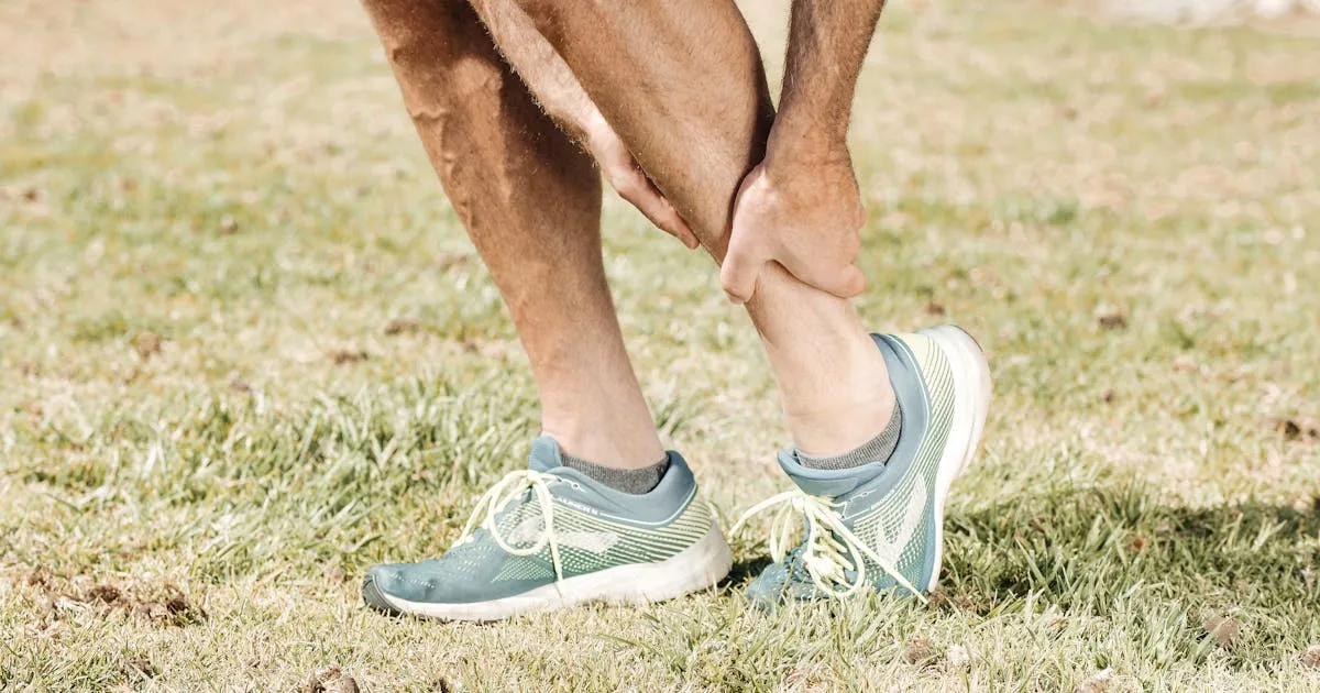
The hallmark symptom of PAES is exercise-induced claudication — a cramping, aching, or tightening sensation in the calf (and sometimes the foot) that comes on predictably during exertion and resolves within minutes of stopping activity. Patients often describe it as a “dead leg” or the feeling that the calf is being squeezed in a vise.
Key features that distinguish PAES from other causes of leg pain include:
- Onset during specific activities: Running (especially uphill or at speed), cycling, and swimming with kick strokes are common triggers. Walking may be less provocative than running.
- Rapid resolution with rest: Unlike musculoskeletal injuries, the pain typically disappears within 2–10 minutes of stopping exercise.
- Young age and high fitness level: According to available data, the majority of patients are between 15 and 40 years old and physically active, and the condition is generally reported in the literature as notably prevalent among military recruits undergoing intense physical training.
- Absent or diminished pulses: During or immediately after exercise, the pulses in the foot (dorsalis pedis and posterior tibial arteries) may be weak or undetectable — a critical clinical sign.
- Cold, pale foot during exertion: Reduced arterial flow can cause pallor and coolness in the foot during an episode.
- Paresthesia: Tingling or numbness in the foot during exercise may occur due to ischemia (insufficient blood supply) affecting nerve function.
In advanced or long-standing cases, repeated trauma to the arterial wall from chronic compression can cause post-stenotic dilatation (widening of the artery just beyond the compression point), thrombosis (blood clot formation), or even aneurysm (abnormal bulging of the arterial wall) — all serious complications that can threaten limb viability.
Diagnosis: What to Expect
Diagnosing PAES requires a high index of clinical suspicion combined with targeted investigations. If you present to a vascular specialist with exertional calf pain, here is what the diagnostic workup typically involves:
Clinical Examination
The vascular specialist will assess pulses at rest and, crucially, after exercise or during provocative maneuvers (active plantar flexion against resistance, or passive dorsiflexion). Disappearance of the foot pulse during these maneuvers is a strong indicator of PAES, though it can also occur in a small percentage of asymptomatic individuals.
Ankle-Brachial Index (ABI)
The ankle-brachial index (a ratio comparing blood pressure at the ankle to blood pressure at the arm) is measured at rest and after a standardized exercise protocol (typically treadmill walking). A significant drop in ABI after exercise — while resting values are normal — is characteristic of PAES and helps differentiate it from peripheral arterial disease (PAD) caused by atherosclerosis (plaque buildup in arteries).
Duplex Ultrasound
Duplex ultrasound (a combined imaging and blood-flow measurement scan) can visualize the popliteal artery and detect compression in real time during provocative positions. It is noninvasive, widely available, and an excellent first-line imaging tool.
MRI and MR Angiography
Magnetic resonance imaging (MRI) and MR angiography (MRA — imaging of the blood vessels using MRI) provide detailed anatomical information about the relationship between the popliteal artery and surrounding muscles. MRI is particularly valuable for identifying the muscular or fibrous band responsible for compression and for planning surgery.
CT Angiography
CT angiography (CTA — X-ray-based detailed imaging of blood vessels) offers excellent spatial resolution and is often used when surgical planning requires precise arterial mapping, or when MRI is not available.
Conventional Angiography
Reserved for complex cases or when endovascular (inside the blood vessel) intervention is being considered, conventional angiography remains the gold standard for depicting arterial anatomy and the degree of compression.
Treatment: From Conservative Care to Surgery
The treatment of PAES depends on whether the condition is functional or anatomical, the severity of symptoms, and the presence of arterial complications.
| Approach | Indication | Details |
|---|---|---|
| Activity modification | Mild functional PAES, early presentation | Temporary reduction in training load; rarely curative alone |
| Surgical decompression | Anatomical PAES, symptomatic functional PAES | Division of the compressing muscle or fibrous band; first-line definitive treatment |
| Arterial reconstruction | PAES with arterial damage (stenosis, aneurysm, thrombosis) | Bypass grafting or patch angioplasty following decompression |
| Endovascular treatment | Selected cases with localized stenosis or thrombosis | Angioplasty ± stenting; generally not recommended as sole treatment |
| Thrombolysis | Acute thrombosis complicating PAES | Clot-dissolving medication to restore flow before definitive surgery |
Surgical decompression — releasing the compressing structure — is the cornerstone of treatment for symptomatic PAES. When performed before irreversible arterial damage occurs, outcomes are excellent. Turnipseed’s 2009 work on functional PAES has been cited in connection with the observation that misdiagnosis may lead to inappropriate interventions (such as fasciotomy for presumed compartment syndrome) that fail to address the true problem, underscoring the importance of accurate diagnosis before any treatment is undertaken. Note: this specific claim should be verified against the original abstract before further attribution.
For patients with established arterial damage — such as a stenosis (narrowing), aneurysm, or occlusion (complete blockage) — surgical decompression is combined with arterial reconstruction, most commonly using a segment of the patient’s own saphenous vein (the long vein running down the inner thigh and calf) as a bypass conduit.
Explore our overview of vascular treatments for more on arterial and venous surgical options.
Prevention: Can PAES Be Avoided?
True anatomical PAES — caused by a congenital (present from birth) anomaly in the artery’s course or muscle position — cannot be prevented, as it is a structural variant. However, several practical considerations apply:
- Early recognition matters most: The best “prevention” of serious complications is early diagnosis. Young athletes with reproducible exertional calf pain that resolves with rest should be evaluated by a vascular specialist, not simply reassured that it is a muscular problem.
- Avoid overtraining in the context of symptoms: In functional PAES, excessive hypertrophy of the gastrocnemius and soleus muscles (the two main calf muscles) may worsen dynamic compression. Modifying training load under medical supervision may slow progression.
- Do not ignore foot pulse changes: If you or a coach notice that the foot becomes cold or pale during intense exercise, this is a vascular warning sign requiring urgent medical assessment.
- Screening in high-risk populations: Some military training programs and elite athletics settings have incorporated vascular screening for recruits with exertional leg symptoms; however, the extent of this practice and the evidence base supporting it vary, and no specific citation is available to confirm how widespread such protocols are.
When to See a Doctor
Consult your physician or a vascular specialist promptly if you experience:
- Calf pain that comes on consistently during exercise and disappears within minutes of rest, especially if you are under 40 and physically active
- Cold, pale, or numb foot during or after exercise
- Absent or weak foot pulses noted during a sports medicine or GP examination
- Sudden severe leg pain at rest — this may indicate acute arterial thrombosis and requires emergency evaluation
- A previous diagnosis of compartment syndrome that did not improve after fasciotomy — functional PAES should be reconsidered
Do not accept “it’s just a muscle problem” if your symptoms follow the classic pattern described above. PAES is treatable, and early intervention prevents permanent arterial damage.
Sources
-
Levien LJ, Veller MG. Popliteal artery entrapment syndrome: more common than previously recognized. Journal of Vascular Surgery. 1999;30(4):587–598. https://doi.org/10.1016/S0741-5214(99)70096-X
-
Darling RC, Buckley CJ, Abbott WM, et al. Intermittent claudication in young athletes: popliteal artery entrapment syndrome. Journal of Trauma. 1974;14(7):543–552. https://doi.org/10.1097/00005373-197407000-00001
-
Turnipseed WD. Functional popliteal artery entrapment syndrome: a poorly understood and often missed diagnosis that is frequently mistreated. Journal of Vascular Surgery. 2009;49(5):1189–1195. https://doi.org/10.1016/j.jvs.2008.12.020
Medical Disclaimer: This article is produced by the Petit Veinard Editorial Board for informational and educational purposes only. It does not constitute medical advice, diagnosis, or a treatment recommendation. Popliteal artery entrapment syndrome is a complex vascular condition requiring evaluation by a qualified physician or vascular specialist. If you are experiencing leg pain, changes in foot circulation, or any of the symptoms described in this article, please consult a healthcare professional promptly. Never delay seeking medical advice on the basis of information read online.
Frequently asked questions
Can a young healthy athlete really have artery problems in the leg?
How is popliteal artery entrapment different from a blood clot in the leg?
What is the treatment for popliteal artery entrapment syndrome and can it be cured?
Petit Veinard Editorial Board
This article was written and reviewed by vascular medicine specialists. Sources: peer-reviewed journals (PubMed), ESVS guidelines, AHA/ACC recommendations, Cochrane Reviews.