Pulmonary Embolism: Recognizing a Life-Threatening Emergency
Pulmonary embolism can be fatal within hours. Learn to recognize the warning signs, understand diagnosis, and know when to call emergency services.
Citable definition: Pulmonary embolism (PE) is an acute (sudden-onset) obstruction of one or more pulmonary arteries — the blood vessels that carry deoxygenated blood from the heart to the lungs — most commonly caused by a blood clot that has migrated from the deep veins of the legs or pelvis. It is a potentially fatal cardiovascular emergency that requires immediate medical evaluation.
What Is Pulmonary Embolism?
Pulmonary embolism sits at the most dangerous end of a disease spectrum known as venous thromboembolism (VTE) — a term that groups together deep vein thrombosis (DVT, a clot in a deep vein, usually in the leg) and PE. In most cases, PE begins silently: a clot forms in a deep vein, grows, and eventually breaks loose. Carried by the venous bloodstream back to the right side of the heart, it is then pumped into the pulmonary circulation, where it lodges and blocks blood flow to part of the lung.
The consequences cascade rapidly. The affected lung tissue can no longer participate in gas exchange (the process by which oxygen enters the blood and carbon dioxide leaves it). The right ventricle — the heart chamber responsible for pumping blood into the lungs — is suddenly forced to work against abnormally high resistance. In severe cases, this right-heart strain can lead to circulatory collapse and cardiac arrest within minutes to hours.
PE is not a rare event. It is estimated to affect approximately 1–2 per 1,000 people per year in Western populations and remains one of the leading causes of preventable in-hospital death. Understanding the biology helps explain why: platelets (tiny blood cells involved in clotting) play a central role alongside clotting proteins in the formation and propagation of venous thrombi. A 2025 review in Blood Reviews highlighted the critical and underappreciated role of platelets in VTE pathogenesis and noted that emerging antiplatelet strategies may complement existing anticoagulation therapies in the future (Liu S et al., Blood Reviews, 2025, PMID: 40382294).
For a broader introduction to venous disease, visit our veins section.
Symptoms — Warning Signs Organized by Frequency
PE is notorious for its variable and sometimes deceptive presentation. Symptoms range from mild and non-specific to dramatic and life-threatening. This variability is one reason the diagnosis is frequently delayed.
Most Common Symptoms
- Sudden shortness of breath (dyspnea): The most frequent presenting symptom, occurring in the majority of patients. It may appear at rest or with minimal exertion.
- Pleuritic chest pain: A sharp, stabbing pain that worsens with deep breathing or coughing, caused by irritation of the pleura (the membrane lining the lungs and chest wall).
- Rapid heart rate (tachycardia): The heart accelerates to compensate for impaired oxygen delivery.
- Cough: Often dry; occasionally productive of blood-tinged sputum (hemoptysis), which suggests pulmonary infarction (death of lung tissue).
Less Common but Clinically Important
- Leg swelling, redness, or pain: Signs of an underlying DVT, present in roughly 50% of PE cases.
- Low-grade fever: Can mimic infection and delay diagnosis.
- Anxiety and a sense of impending doom: Frequently reported by patients and not to be dismissed.
The Atypical Presentation — Syncope
Perhaps the most alarming atypical presentation is syncope (a sudden, transient loss of consciousness). A 2025 case report published in Cureus described a patient who presented with syncope as the sole initial symptom of a hemodynamically significant PE — and notably did so without the expected drop in blood oxygen levels (hypoxemia). The authors highlighted that severe PE can present without hypoxemia, underscoring the diagnostic challenge of atypical presentations (Wery L, Cureus, 2025, PMID: 40755597).
Key message: There is no “typical” PE patient. When in doubt, seek emergency care immediately.
Diagnosis — What to Expect in the Emergency Department
Diagnosing PE is a structured process designed to avoid both under-diagnosis (missing a life-threatening clot) and over-diagnosis (exposing patients to unnecessary radiation and anticoagulation risks).
Step 1 — Clinical Probability Assessment
Emergency physicians use validated scoring tools to estimate how likely PE is before ordering tests. The most widely used is the Wells score, which assigns points for clinical features such as signs of DVT, heart rate above 100 bpm, immobilization, and the absence of an alternative diagnosis. The Wells score was originally developed and validated by Wells et al. in a series of studies published between 1998 and 2003. Separately, Kline et al. developed the Pulmonary Embolism Rule-out Criteria (PERC rule), a clinical decision tool designed to identify low-risk patients in whom further diagnostic testing for PE may be safely avoided, according to available data.
Step 2 — D-Dimer Blood Test
D-dimer is a protein fragment released when a blood clot breaks down. A negative D-dimer result in a low-probability patient effectively rules out PE without further imaging. However, D-dimer is non-specific — it is elevated in many other conditions (infection, pregnancy, cancer, recent surgery) — so a positive result alone does not confirm PE.
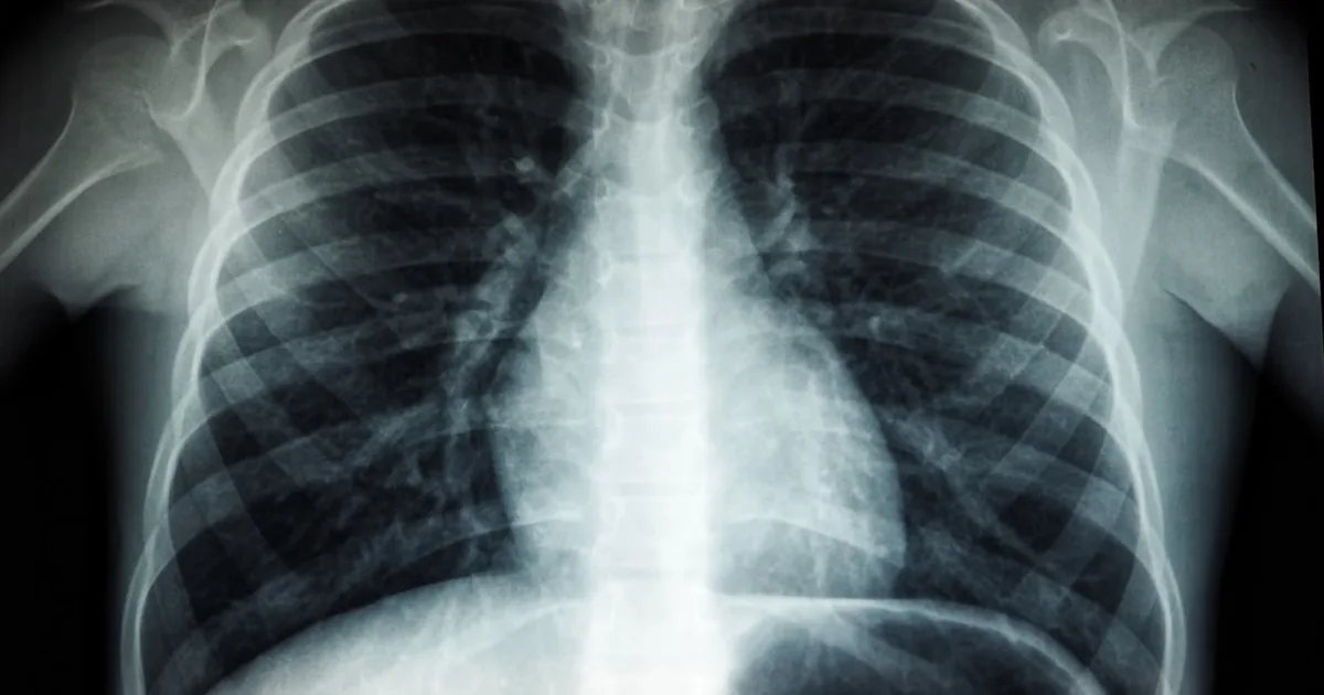
Step 3 — CT Pulmonary Angiography (CTPA)
CTPA — a specialized CT scan in which contrast dye is injected to visualize the pulmonary arteries — is the gold-standard imaging test for PE. It offers high sensitivity and specificity for detecting clots in the pulmonary arteries, and its appropriate use must be guided by clinical context and pre-test probability assessment.
Step 4 — Echocardiography and Severity Assessment
In hemodynamically unstable patients (those in shock or near-collapse), bedside echocardiography (ultrasound of the heart) can rapidly confirm right-heart strain and guide emergency treatment. It is also used to risk-stratify patients once PE is confirmed, helping to distinguish between low-risk, intermediate-risk, and high-risk (massive) PE.
Differential Diagnoses to Rule Out
Chest pain and breathlessness have many causes. Emergency physicians must also consider conditions such as acute coronary syndrome (heart attack), pneumothorax (collapsed lung), aortic dissection, and — importantly — rarer diagnoses. For completeness, it is worth noting that esophageal perforation (Boerhaave syndrome), a surgical emergency presenting with chest pain, is one differential that must be considered in specific clinical contexts (Tayyib M et al., Cureus, 2025, PMID: 41268039).
Treatment — From Anticoagulation to Emergency Intervention
| Severity | Hemodynamic Status | Primary Treatment |
|---|---|---|
| Low-risk PE | Stable, no right-heart strain | Anticoagulation alone; outpatient possible |
| Intermediate-risk PE | Stable, but right-heart strain on imaging/biomarkers | Anticoagulation; close monitoring; consider thrombolysis |
| High-risk (massive) PE | Unstable — shock or cardiac arrest | Systemic thrombolysis or surgical/catheter embolectomy |
Anticoagulation (Blood Thinners)
Anticoagulation is the cornerstone of PE treatment. It does not dissolve the existing clot but prevents it from growing and allows the body’s own fibrinolytic (clot-dissolving) system to work. Options include:
- Direct oral anticoagulants (DOACs) such as rivaroxaban or apixaban — now the first-line choice in most patients per both European Society of Vascular Surgery (ESVS) and American Heart Association/American College of Cardiology (AHA/ACC) guidelines.
- Low-molecular-weight heparin (LMWH) — injected subcutaneously, often used as a bridge or in patients with cancer-associated VTE.
- Unfractionated heparin (UFH) — intravenous, preferred in high-risk PE where rapid reversal may be needed.
Thrombolysis
Systemic thrombolysis (administration of a clot-dissolving drug, typically alteplase, through a vein) is reserved for high-risk PE with cardiovascular collapse. It rapidly restores blood flow but carries a significant risk of major bleeding, including intracranial hemorrhage (bleeding in the brain). A comprehensive overview of acute PE management — including the role of thrombolysis — was provided by Tapson VF in a 2005 review published in Cardiology Clinics (Tapson VF, Cardiology Clinics, 2005).
Catheter-Directed Therapy and Surgical Embolectomy
In specialized centers, catheter-directed thrombolysis (delivering the drug directly into the clot via a catheter) or surgical embolectomy (physically removing the clot) may be considered for patients who cannot receive systemic thrombolysis or do not respond to it.
For a full overview of vascular treatment options, explore our treatments section.
Prevention — Actionable Daily Steps
PE is largely preventable. Risk reduction strategies apply both to the general population and to high-risk individuals (post-surgical patients, long-haul travelers, those with cancer or clotting disorders).
- Stay mobile: Prolonged immobility is a major risk factor. On long flights or car journeys, stand up and walk every 1–2 hours. Perform calf-pump exercises (flexing and extending the ankle) when seated.
- Hydration: Dehydration thickens the blood. Drink water consistently, especially during travel and illness.
- Compression stockings: Graduated compression hosiery reduces venous stasis (sluggish blood flow) in the legs. Recommended for travelers at moderate-to-high VTE risk.
- Prescribed anticoagulation: Patients undergoing major orthopedic or abdominal surgery are routinely prescribed prophylactic (preventive) anticoagulation — do not stop it without medical advice.
- Know your risk factors: Obesity, smoking, oral contraceptives, pregnancy, personal or family history of VTE, and inherited clotting disorders (thrombophilia) all increase PE risk. Discuss these with your physician.
More evidence-based prevention strategies are available in our prevention section.
When to See a Doctor — Clear Criteria for Seeking Medical Attention
Call emergency services (112 in Europe, 911 in the US) immediately if you experience:
- Sudden unexplained shortness of breath
- Chest pain, especially if it worsens with breathing
- Fainting or near-fainting without an obvious cause
- Coughing up blood
- Rapid or irregular heartbeat combined with any of the above
Schedule an urgent appointment with your physician or vascular specialist if you have:
- A swollen, red, or painful leg — even without chest symptoms — as this may indicate DVT that could progress to PE
- Known risk factors for VTE and are planning surgery, long travel, or pregnancy
- Recently completed anticoagulation treatment and are experiencing new symptoms
Do not wait and see. PE can be fatal within the first hours of onset. Early treatment dramatically improves outcomes.
Sources
-
Liu S, Shen Y, Chen J, et al. The critical role of platelets in venous thromboembolism: Pathogenesis, clinical status, and emerging therapeutic strategies. Blood Reviews. 2025 Nov;74:101302. DOI: 10.1016/j.blre.2025.101302 | PMID: 40382294
-
Wery L. Syncope as the Initial Presentation of Severe Pulmonary Embolism Without Hypoxemia: A Clinical Case. Cureus. 2025 Jul;17(7):e87159. DOI: 10.7759/cureus.87159 | PMID: 40755597
-
Tayyib M, Latif MF, Sakr A, Mahmoud A. Chest Pain After Vomiting: Recognizing Boerhaave Syndrome in the Emergency Department. Cureus. 2025 Nov;17(11):e97219. DOI: 10.7759/cureus.97219 | PMID: 41268039
-
Tapson VF. Acute pulmonary embolism. Cardiology Clinics. 2005;23(1):1–14.
⚠️ Medical Disclaimer
This article is produced by the Petit Veinard Editorial Board for informational and educational purposes only. It does not constitute medical advice, diagnosis, or a treatment recommendation. The information presented reflects published scientific literature and established clinical guidelines available at the time of writing and may not apply to every individual situation. Always consult a qualified physician or vascular specialist before making any decisions about your health. In a medical emergency, call your local emergency number immediately. Petit Veinard is an independent media outlet and has no financial relationships with pharmaceutical or medical device companies.
Frequently asked questions
What are the first signs of a pulmonary embolism I should watch for?
Can a blood clot in the leg travel to the lungs?
How is pulmonary embolism diagnosed in the emergency department?
Petit Veinard Editorial Board
This article was written and reviewed by vascular medicine specialists. Sources: peer-reviewed journals (PubMed), ESVS guidelines, AHA/ACC recommendations, Cochrane Reviews.